

Hydatid Disease (Part V), wherein we finally explain why it makes you sick.
It takes years for symptoms to show up. Oh, but when they do.... The Liver The growing cyst puts pressure on the liver, producing symptoms of obstructive jaundice and abdominal pain. If the cyst ruptures into the biliary tree (the collection system of bile in the liver), severe, possibly life-threatening inflammation along with intense pain and jaundice. The most serious problem, though, is the rupture of the cyst. This occurs in 50-90% of the cases. It can cause anaphylactic


Hydatid Disease (Part IV)
The larval form of E. granulosus manifests itself as a cyst, called a hydatid cyst. The cyst is actually not a single larva, but a fluid filled balloon of tissue filled with large numbers of tiny larval forms, called protoscolices (sing. protoscolex). Each protoscolex has the potential, if ingested by a dog, of becoming an adult tapeworm in the intestines of a dog, where the cycle can start again. Note that dogs carry these worms without problems or symptoms. You cannot tell

Hydatid Disease (Part III)
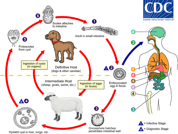
Above is the life cycle of E. granulosus. Life is fine for all concerned if the pattern follows the red lines, However, if there's the detour to the blue lines--the accidental ingestion of an egg by a human instead of a sheep--then consequences ensue. Notice that the egg is shed in feces. Dog poop. Since this parasite is transmitted to humans via the all-too-common route of fecal-oral, good hygiene ( in theory) should prevent this. We all know that hand-washing, the most impo